UCLA researchers have developed an innovative approach to diagnosing cancer. This research has excellent commercial applications and is already in the process of being translated into a novel technology by a Los Angeles start up. The new technique allows researchers to very accurately diagnose cancers by observing the physical characteristics of cells in bodily fluids.
This approach is particularly noteworthy because it would allow scientists to diagnose the presence of cancerous cells without having to remove them from the body fluid in which they are present. The isolation of cells for testing is one of the most costly and time consuming aspects of cancer diagnostics. These isolating processes can involve using complex dying or molecular marking processes that are often not accurate.
The technique developed by the UCLA researchers bypasses all these complex aspects of sample preparation and allows scientists to diagnose cancerous cells in the body fluid in which they are found. The technique uses a form of cytometry to take images of cells as they flow through tiny fluid channels. These images are then analyzed to determine if the cell is cancerous or healthy.

(Image of Flow Cytometer)
In particular, the characteristics that the cytometer is testing has to do with how the cell responds to being “squeezed” by slight pressure in the fluid that surrounds it. This type of cytometry is known as “deformability cytometry.” By taking images of how cells respond to pressure, scientists can glean clues to their internal structure and determine whether they are cancerous or not. Cells that have become cancerous have a very different internal structure from healthy cells and thus deform in a much different way in the cytometry process.
Since this technique is imaging and then analyzing cells as they flow in a fluid, rather than isolating individual cells for analysis, it has a much higher capacity than previous techniques. The cytometry technique can analyze 1,000 cells per second. This not only allows scientists to more quickly test for cancer, it is also much more accurate. By allowing scientists to analyze a much larger sample size with more detail, they can get a better sense of the variations and predominance of the cancer in the patient. This new cytometry technique was able to identify cancer in several patients that could not be diagnosed using previous techniques.
This cytometry technique is continuing to be developed and refined by a team of UCLA researchers. As Dino Di Carlo, co-principal investigator of this research project and an associate professor of bioengineering at UCLA’s Henry Samueli School of Engineering and Applied Science said, "Building off of these results, we are starting studies with many more patients to determine if this could be a cost-effective diagnostic tool and provide even more detailed information about cancer origin. It could help to reduce laboratory workload and accelerate diagnosis, as well as offer doctors a new way to improve clinical decision-making."
Jianyu Rao, the other co-principal investigator and a professor of pathology and laboratory medicine at UCLA’s David Geffen School of Medicine summarized the potential benefits of this research. He said, "First, it may increase diagnostic accuracy for the detection of cancer cells in body fluid samples. Second, it may provide a method of initial screening for cancer in body fluid samples in places with limited resources or a lack of experienced cytologists. Third, it may provide a test to determine the drug sensitivity of cancer cells."

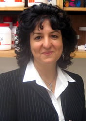
(Image of Jianyu Rao from UCLA)
This research has already spawned a new startup dedicated to commercializing and translating this technique into a product that could directly help physicians in diagnosing cancer. The new startup, called Cytovale, has already received a competive grant from Breakout Labs. This foundation focuses on funding groundbreaking technologies with innovative applications.
This research is an example of the vibrant life science community in Los Angeles and shows how new research can have wide reaching affects. This research project has directly led to the creation of a new startup that will vitalize the life science industry in Los Angeles. This is one example of how research at UCLA spills over into the broader community to create the innovative research culture of Los Angeles life science research.
Biotechnology Calendar, Inc. will be holding its Semiannual UCLA Biotechnology Vendor Showcase™ expo event on April 4, 2014. This life science marketing event is an excellent opportunity for life scientists and lab equipment specialists to take advantage of the unique research culture around UCLA and network around latest developing lab technologies.
For information on exhibiting at this life science marketing event and a 1-page funding report, click on the button below.


