
To understand the complex working of biological molecules, sharp images are the key. Electron microscopes do a good job when it comes to inorganic compounds but don’t lend quite as easily to organic molecules, especially if you want to view them alive and in action. Researchers at the University of Illinois, Chicago have developed a new method for imaging biomolecules that solves many of the current problems in the field.
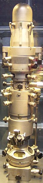 (An electron microscope pictured at right, courtesy Wikimedia Commons and Stahlkocher)
Electron microscopes view samples in a vacuum. This is satisfactory for the viewing of many atoms and molecules, but is rather inadequate in the case of biomolecules, which naturally exist in an aqueous environment. Current solutions to this problem involve placing biological samples in a container filled with liquid; however, to withstand the vacuum, the walls of this container must be relatively thick, making for fuzzier images.
 The UIC team presented a new approach: encapsulate the liquid instead in graphene. Graphene layers are only one atom thick, so the feat of keeping liquid safely inside means the world in terms of viewing clarity. In the words of Robert Klie (left), senior investigator of the study, “It’s like the difference between looking through Saran Wrap and thick crystal.”
Another significant advantage of using graphene is the protection from radiation that it grants. Electron microscopes shoot electron beams at their sample in order to generate their images. This can destroy biological samples, so the intensity is turned way down, with the trade off again being loss of clarity. The graphene solves this problem as well, says Klie, because it can “conduct away both the heat and the electrons generated as the electron microscope’s beam passes through the sample,” as he puts it in a UIC news release.
This new method will be especially useful in the imaging of ferritin, a biomolecule that regulates iron in organisms. It’s well known that defects in ferritin lead to several disorders and diseases, but not exactly how or why. Using graphene will allow bioresearchers to image the core of the molecule, which has not yet been done, to fully understand it at the molecular level. Of course, the scope of this achievement goes beyond one particular molecule; Klie believes it will “open up analysis of biological and other difficult to image samples to almost anyone with an electron microscope.”
This work was funded by grants from the NSF and MTU. To obtain more detailed information regarding funding, research, and development at UIC, read Biotechnology Calendar’s complimentary University of Illinois, Chicago Funding and Vendor Statistics report.

Biotechnology Calendar, Inc. makes an appearance at the University of Illinois Chicago campus each year for its Chicago BioResearch Product Faire™, held next on May 01, 2014. If you're in the area, we will also be hosting the Urbana-Champaign Bioresearch Product Faire™ on the UIUC campus the day before, April 31, 2014. Biotechnology Calendar, Inc. is a full service event company that has produced on-campus, life science research trade shows nationwide for the past 20 years. If you are a university researcher or a laboratory product vendor, consider attending one of our on-campus trade shows: here is our 2014 schedule of events.

